Ultrasound Machines: Exploring Emerging Trends in Diagnostic Technology
Download Now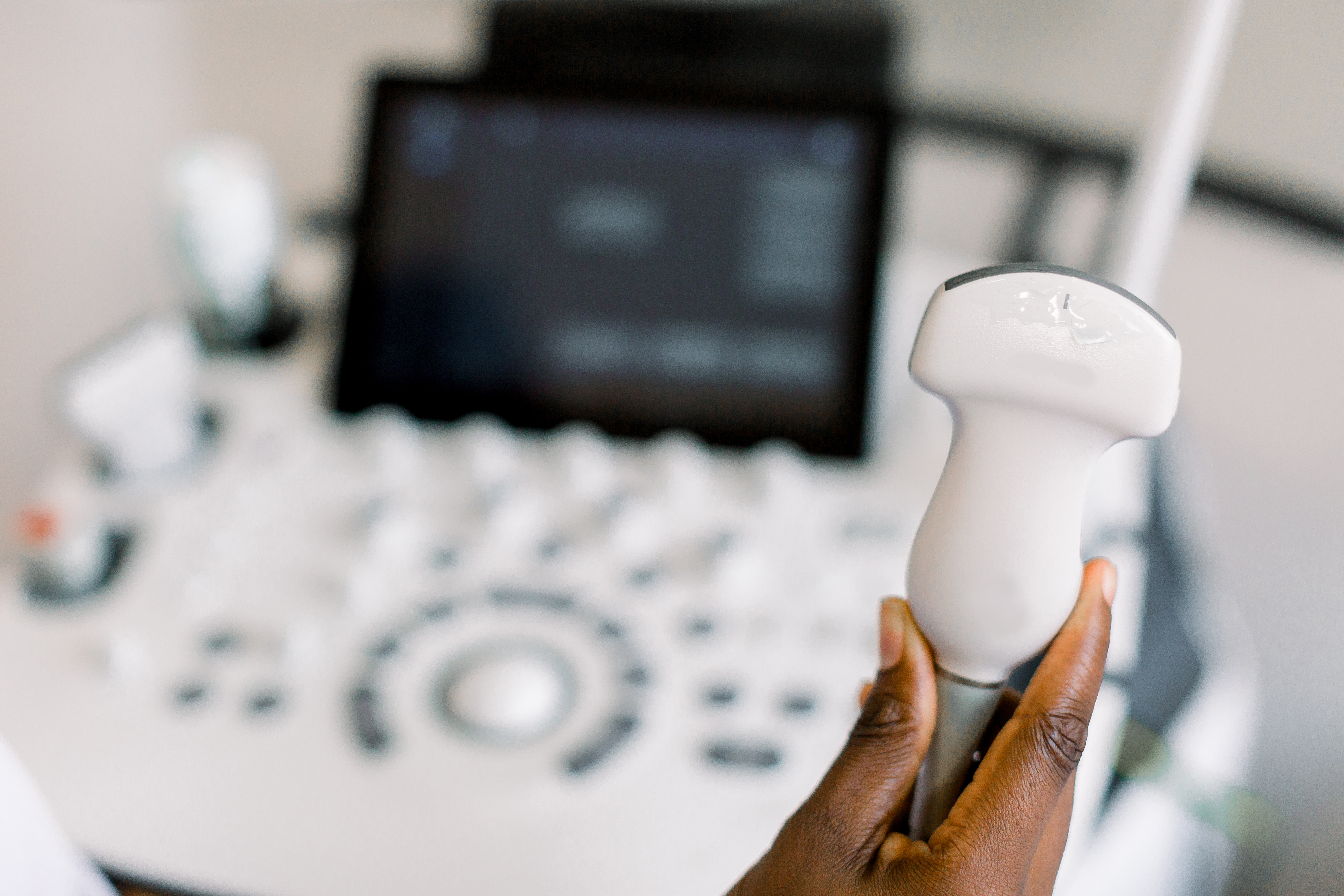
Table Of Contents
With its initial development in the late 1950s, ultrasound technology revolutionized medical diagnostics - so much so that today, it is impossible to imagine practicing in fields like obstetrics and gynecology without one of the many ultrasound machines on the market.
However, thanks to recent advances in ultrasound research, these machines are capable of much more than a non-invasive and cost-effective method of visualizing body structures.
High-definition imaging, AI integration, and 3D capabilities are enhancing diagnostic precision and patient care while also boosting efficiency within clinical settings, facilitating team collaboration, and alleviating the healthcare burden. This guide will cover everything needed for sonographers and specialists to stay up to date with the latest innovations and choose the best ultrasound machine for their practice.
Want a copy for later? Download the full guide here.
The Physics Of Ultrasound Waves
Ultrasound machines use high-frequency sound waves (in the megahertz range, above the threshold of human hearing) to create images of the body’s internal structures.
They leverage a transducer, or probe, which emits sound waves that travel into the body and bounce off tissues and organs.
When the sound waves encounter different tissues, they are either reflected, refracted, or attenuated:
Reflection
A portion of the wave reflects back to the transducer when ultrasound waves encounter a boundary - or a surface where two different materials with distinct acoustic properties meet, like muscle and bone. This principle is used in imaging soft tissues and detecting abnormalities like tumors.
Refraction
This occurs when ultrasound waves pass through media of different densities at an angle, which causes them to change direction before being captured by the probes. This principle can be used to visualize the heart through the rib cage.
Attenuation
When ultrasound waves travel through tissues, their intensity diminishes due to physics principles like absorption and scattering, indicating malformations and abnormal structures.
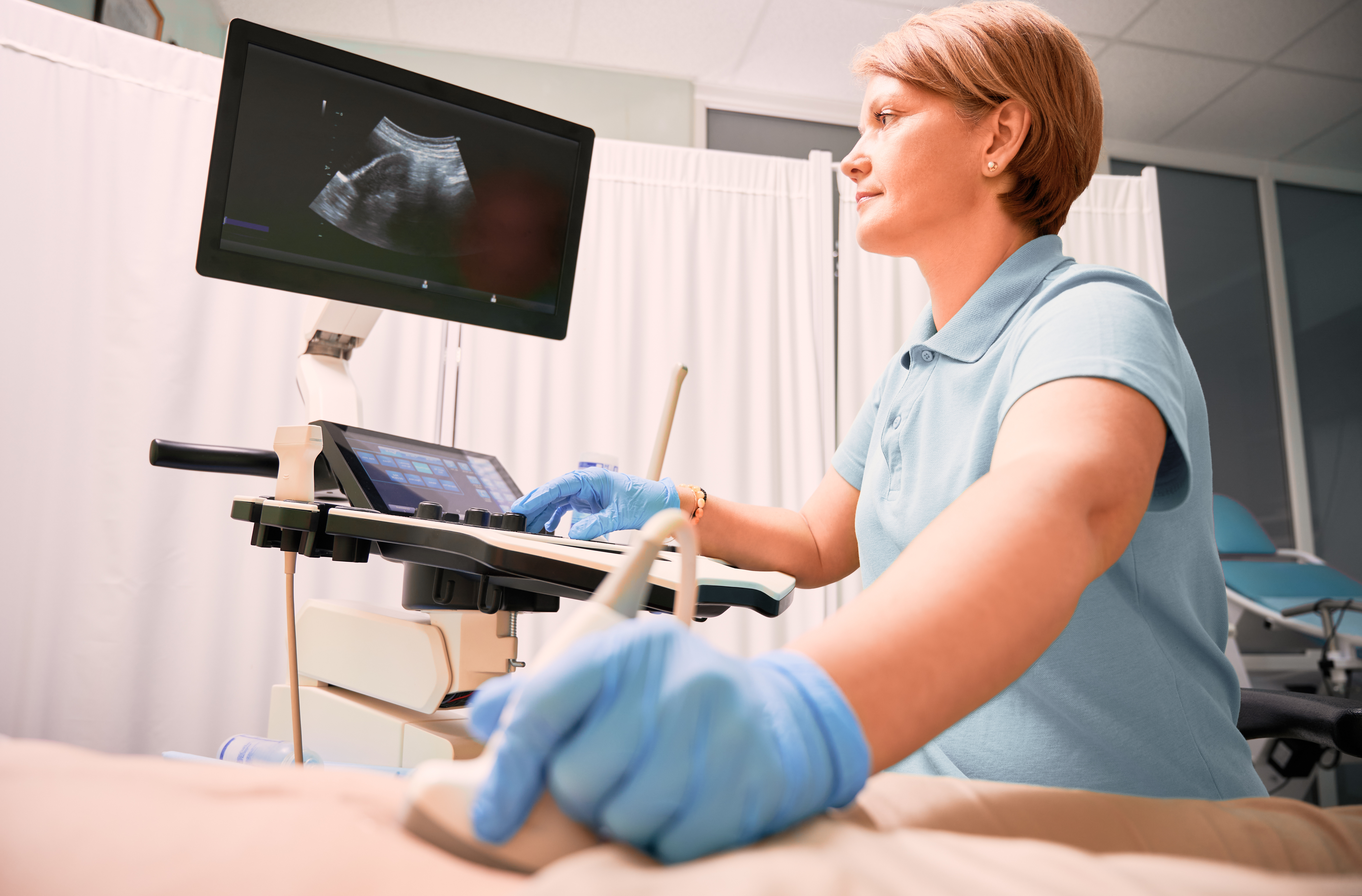
The transducer then captures these echoes and sends them to a computer, which processes and converts them into real-time visual images on a monitor. Sonographers can then read and interpret these images.
Although the principles at the core of ultrasound technologies have remained stable since its inception, new and emerging innovations have enhanced the accuracy, clarity, clinical applications, and ease of use of ultrasound tools. Today’s physicians can access a range of imaging standards and types, including:
- 2D ultrasound
Standard imaging providing two-dimensional images. - 3D ultrasound
Captures volume data to offer more detailed views of structures and from multiple angles. - 4D ultrasound
Offers live stream of 3D images, creating a real-time video of internal structures. - Doppler ultrasound
Measures and visualizes blood flow within vessels, detecting blockages or clots. It allows for the use of ultrasound in fields like vascular medicine. - Tissue harmonic imaging
Enhances image quality by using harmonic waves generated within the body, which reduce “noise.” - Contrast-enhanced ultrasound
Improves the visualization of blood flow and tissues through contrast agents. - Speckle reduction imaging
Reduces speckle noise to produce clearer, more accurate images.
Traditional And Emerging Clinical Applications Of Ultrasound Machines
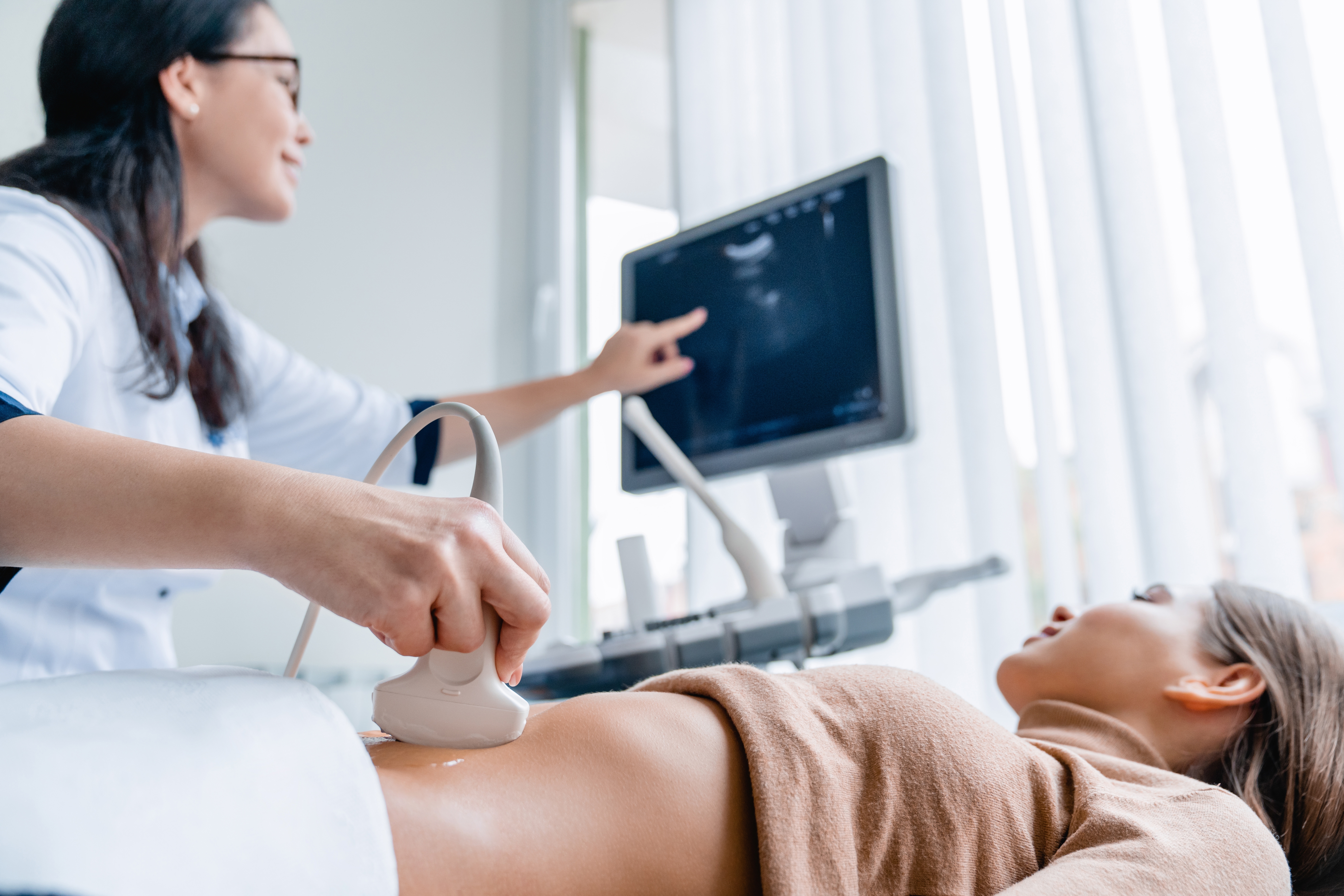
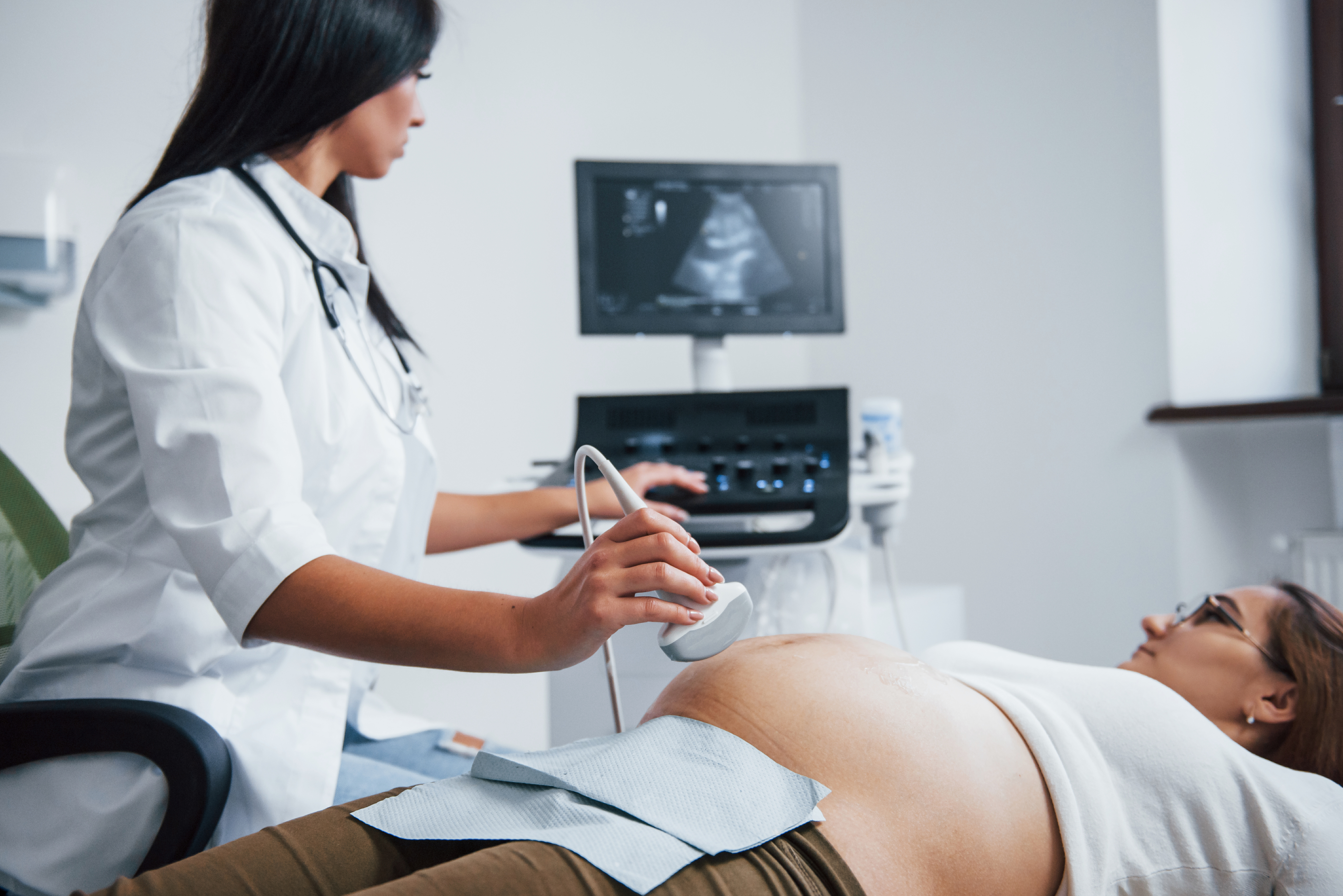
Ultrasound machines are commonly associated with maternal-fetal medicine (MFM), or pregnancy care, for tasks like the detection of pregnancy and fetal anomalies and monitoring high-risk pregnancies.
While ultrasound machines remain a cornerstone in obstetrics, their clinical applications extend well beyond this field. For decades, these machines have been vital in fields like cardiology for echocardiograms, internal medicine for diagnosing liver and kidney conditions, and guiding various biopsies and fluid drainage procedures.

Recent innovations have broadened the scope of ultrasound technology even further, making it a viable diagnostic and therapeutic alternative in fields previously dominated by other diagnostic tools. These included cardiology and musculoskeletal imaging, which used to rely on cumbersome CT and MRI scans - technologies with downsides such as limited accessibility, high costs, and radiation exposure risks.
State-of-the-art ultrasound machines today can produce real-time, high-quality images of soft tissues and integrate technologies like automation, AI, and machine learning.
These developments have made them a more affordable, lower-risk, and ultimately indispensable tool in various fields, including vascular medicine, gastroenterology, musculoskeletal imaging, and more. Let’s explore these applications further below.
Medical Diagnostics
Ultrasound technology plays a critical role in medical diagnostics. It offers a non-invasive, real-time imaging method that helps physicians evaluate and manage various health conditions, such as identifying abnormal growths and tumors, assessing the progression of degenerative conditions, and monitoring fetal development.
Some of the diagnostic applications of ultrasound technology include:
Obstetrics and gynecology
Since its inception, ultrasound has been a critical ally in pregnancy care. It helps monitor fetal development, assess the placenta, and detect anomalies such
as ectopic pregnancies and congenital defects. Advanced 3D and 4D imaging allow for more
detailed visualization, enabling early intervention and better outcomes for mothers and babies. Today, second-trimester ultrasound is the gold standard for detecting structural anomalies, while the use of this technology in the first trimester is increasing.
Cardiology
In cardiology, ultrasound waves, or echocardiograms, can visualize heart structures, assess the heart’s function, and diagnose conditions like heart valve disorders and cardiomyopathies. In particular, tissue Doppler imaging has been used to evaluate diastolic heart failure, a common complication in post- COVID-19 patient care.
Oncology
Ultrasound aids in identifying and monitoring various tumors and abnormal tissue growth, particularly in the breast, liver, and thyroid.
Vascular
By reflecting off moving blood cells, Doppler ultrasound provides the accuracy necessary to assess blood flow in arteries and veins and detect vascular diseases like deep vein thrombosis and atherosclerosis.
Urology
In urology, ultrasound is used to diagnose kidney stones, bladder tumors, and prostate issues. Because it is non-invasive and quick, ultrasound is usually a first-line imaging technique that helps address time- sensitive health conditions.
Gastroenterology
In gastroenterology, endoscopic ultrasound (EUS) provides detailed images of the digestive tract and surrounding tissues, helping diagnose conditions like pancreatitis and gastrointestinal tumors.
Integrating advanced features like 3D imaging and AI has further enhanced ultrasound’s diagnostic capacity, making it a vital tool across medical specialties beyond the ones seen above.
Therapeutic And Surgical Uses
Ultrasound technology isn’t limited to diagnostics - it also plays a pivotal role in delivering therapies and guiding surgery in a minimally invasive way. Some of the therapeutic and surgical uses of ultrasound tools include:
Focused ultrasound surgery (FUS)
FUS uses targeted ultrasound waves to treat tissues without the need for incisions. For example, high- intensity focused ultrasound (HIFU) can deliver targeted heat to specific areas of the body. This technology can ablate tumors or uterine fibroids without affecting nearby healthy tissues. This method reduces recovery times and has fewer side effects than traditional surgeries. MRI-guided HIFU is now also used in treating tremors (by heating brain tissue and blocking the signals that cause tremors).
Ultrasound-guided interventions
Ultrasound can also guide other procedures, enhance targeted interventions, and boost accuracy. For example, real-time imaging aids physicians when delivering injections by helping them visualize
needle placement from the point of entry. Typical applications include guiding stem cell injections and platelet-rich plasma (PRP) therapy in regenerative orthobiologics.
Given the role played by ultrasound in these emerging therapies, it is easy to see why this tool is likely to become only more indispensable over the next few years.
8 Recent Advances In Ultrasound Technology
Over the past years, ultrasound technology has developed at an unprecedented rate, driven by modern healthcare research and the emergence of tools like AI, automation, and machine learning.
These developments have enabled ultrasound machines to become essential in most healthcare practices, allowing them to manage the increasing patient load without compromising standards of care.
Beyond improving practice management, ultrasound machines’ faster, less invasive diagnostic capabilities can also help improve patient outcomes. For example, this speed and accuracy can be life-saving in vascular medicine, where patients often move quickly from ultrasound exams to CT scans or surgery.
Nonetheless, with ultrasound machines developing faster than ever, it can be challenging for sonographers and practice managers to keep up with the latest innovations and standards.
Below are some of the technologies to look for in a cutting-edge ultrasound machine - but two aspects should never be overlooked:
-
Technologies emerge, change, and possibly fade quickly, and it’s essential for physicians to stay up to date with the latest changes.
-
Your practice’s specific needs may call for certain features over others, an aspect we’ll discuss in more detail below.
1. Miniaturization and Portability
In recent years, advances in miniaturization and portability in ultrasound technology have revolutionized how diagnostic imaging is approached. Where previously, patients had to be brought into a specialized room for ultrasound tests, today, handheld and portable machines are meeting the needs of patients where they are.
In most settings, handheld devices have made ultrasound accessible beyond traditional imaging centers, enhancing on-site accessibility. These devices, usually ergonomic, lightweight, and about the size of a smartphone, connect to tablets or smartphones, offering high-quality imaging on a mobile platform.
Moreover, equipped with multiple probes, they can handle various diagnostic needs—such as diagnosing volume overload in an emergency care patient —without requiring device changes.
Impact on Point-of-Care Diagnostics
Portable ultrasound machines empower healthcare providers to perform diagnostic imaging at the patient’s bedside—an innovation known as point-of-care ultrasound (POCUS). This enhances patient comfort
and improves diagnostic speed and accuracy in critical settings, such as preliminary assessments in an emergency room or monitoring in intensive care.
The portability of these devices does not impact their accuracy. According to a 2024 study, POCUS demonstrated 96.6% accuracy, making it a highly accurate diagnostic tool for virtually all conditions performed by emergency department physicians.
Examples of Use
One of the most significant advantages of miniaturization and portability is that these features allow ultrasound exams to be performed across a wide range of settings and circumstances:
Crowded diagnostic rooms
Handheld devices can fit in tight spaces in busy hospital environments, allowing continuous and easy access to imaging.
Emergency departments
Rapid diagnostic capabilities at the patient’s bedside are crucial in emergency medicine to make potentially life-saving, well-informed decisions.
Remote locations
Portable devices deliver healthcare in underserved or geographically isolated areas, bridging the gap between rural communities and medical care.
Patients with limited mobility
Bedridden or critically ill patients can also benefit from bedside imaging, which reduces the strain of moving them to other departments, such as radiology wards.
Continuity of care
The availability of ultrasound images across different settings ensures ongoing care without interruption.
2. High Resolution Imaging
When it comes to ultrasound machines, the image resolution refers to how clear the image is and how far it can be zoomed in without losing that clarity.
There are different types of resolution for ultrasounds, including temporal resolution, which measures changes over time, and spatial resolution, which includes axial and lateral resolution, for measuring the size of structures and separations between them.
Spatial Resolution
The spatial resolution of an ultrasound determines its ability to distinguish between different structures. An image with low spatial resolution will appear blurry, making it challenging to identify the spaces between structures within an ultrasound image.
Since movement can negatively impact spatial resolution, patients must stay very still during exams, especially when healthcare professionals are looking for minute details in a scan. Other factors impacting spatial resolution include:
- The size of the field being examined
- The depth of the structures in the scan
- The pulse length
Shallower images with shorter pulses appear clearer since the ultrasonic waves don’t need to travel as far before they return. Smaller view fields are also clearer than wide ones due to the concentration of the waves.
Ultrasound machines with wider transducers may produce higher-resolution images than ones with narrow probes. Machines that operate efficiently at shorter pulse lengths may also show clearer images.
Spatial resolution can be broken down into axial and lateral resolution. The type required depends on the orientation of the scanned structures relative to the ultrasound beam.
An ultrasound’s axial resolution refers to its ability to distinguish separate structures side-by-side when they are oriented parallel to the beam. Machines using a higher frequency and shorter pulse length offer better axial resolution. However, the shorter the pulse, the shallower the image will be.
Axial resolution is typically clearer than lateral. This is because the elements are positioned along the same line as the ultrasound, meaning the beam doesn’t need to be spread so far to distinguish between structures.
Lateral Resolution
Lateral resolution enables a sonographer to distinguish between structures oriented side-by-side when perpendicular to the beam. The gap between the structures needs to be wider than the ultrasound beam being used to separate them. Lateral resolution is usually much lower than axial resolution and is affected by the number of lines per image, the size of the transducer, and how focused the beam is.
Shorter pulses across a smaller field produce the clearest views. However, if you need to examine a wider region or the affected structures require deeper penetration, this will reduce the resolution. In such cases, a higher-end ultrasound machine that can enhance images may be beneficial.
Temporal Resolution
The temporal resolution of an ultrasound image designates the software’s ability to distinguish minuscule changes over time. This is used to identify factors such as movement within an organ structure or the speed and direction of blood flow through veins and arteries. Temporal resolution is determined by the field’s pulse length, depth, and width.
Smaller fields of view and shorter pulse lengths both increase temporal resolution.Ultrasounds with Doppler capabilities rely on temporal resolution to monitor directional movement over time. Doppler ultrasounds are most commonly used to track blood flow. In such uses, blood traveling toward the transducer will appear red, and blood traveling away from the pulses will read as blue.
3. Integration with AI and Machine Learning
One of the most groundbreaking advancements in ultrasound technology is the integration of artificial intelligence and machine learning:
Artificial intelligence (AI)
AI technology allows machines to simulate human intelligence. It can help automatically detect structures and abnormalities during scans, improving diagnosis accuracy and efficiency.
Machine learning (ML)
ML is a subcategory of AI designed to allow systems to learn and improve from experience. ML algorithms analyze ultrasound images in real time, enhancing image quality and helping physicians perform complex diagnostic tasks.
These technologies are pushing the boundaries of what ultrasound machines can do, making them more intelligent, accurate, and efficient tools capable of reducing timelines and human error.
Some of the capabilities of AI and ML include:
Predictive analytics in diagnostics
Predictive analytics are driven by machine learning and AI. By analyzing vast amounts of data, they can predict potential health issues before they become critical.
For instance, AI-enabled ultrasound systems can identify patterns in fetal growth, pointing to potential complications early in pregnancy.
Automated image interpretation
AI algorithms can assess and interpret images in real time, automating the identification and analysis of anatomical structures, anomalies, and malformations.This innovation reduces the likelihood of human error and shortens examination time in critical fields such as
fetal ultrasound. To visualize this benefit, AI can process and compare up to 10 images per second, while a human technician might take several minutes to assess a single image accurately.

4. Accessibility
Advancements in ultrasound technology have dramatically enhanced the accessibility of powerful diagnostic images in terms of cost and availability in low-resource settings.
To see how, it’s enough to look at the high cost and complexity associated with CT scans. These can average from $500 to $3,000, excluding additional expenses for contrast agents, and can be highly time-consuming. On the other hand, an ultrasound for diagnosing conditions like deep vein thrombosis or monitoring vascular health can cost from $200 and $800. Moreover, ultrasound scans are much quicker, can be delivered at the patient’s bedside, and avoid the need for contrast agents, making the process less cumbersome and resource-intensive.
The cost-effectiveness of ultrasound imaging over CT and MRI scans also benefits hospitals that accept large volumes of Medicare and Medicaid patients. By providing faster and more affordable diagnoses, high-quality ultrasound machines can help these hospitals remain profitable without compromising diagnostic and care quality.

5. Innovations in Transducer Technology
As mentioned above, transducer technology is at the heart of ultrasound imaging. Transducers convert electrical signals into sound waves and back into electrical signals after they travel through body tissues, allowing computers to read and plot information.
In the early days of ultrasound scans, traditional transducers were limited in their frequency ranges and resolution, leading to less accurate scans, limited penetration capabilities, and lower-quality images. However, the development of the next generation of high-frequency transducers has revolutionized the field. These technologies include:
- Piezoelectric transducers use piezoelectric crystals to convert electrical signals into sound waves and vice versa.
- Photoacoustic transducers combine laser-induced ultrasound waves to generate images.
- Capacitive micromachined ultrasonic transducers (CMUTs) are made using silicon to produce and detect high-frequency sound waves.
- Ultrasonic transducers: are specialized devices that detect ultrasonic waves, providing high-frequency sound waves beyond human hearing.
6. Use of Contrast Agents
Many high-end features aid in advanced imaging, which provides a clearer picture of a patient’s condition. Advanced features may include contrast ultrasounds, elastography, or 3D/4D ultrasound capabilities.
In particular, contrast-enhanced ultrasounds use a dye inserted into the structure or bloodstream to help identify abnormalities. Not all ultrasounds can distinguish this dye, so this would be considered an advanced feature of some higher-end machines.
Some of the benefits of this innovation - especially when coupled with AI integrations - include:
- High penetration: Provides deeper visibility and more accurate diagnosis, even with significant tissue mass, making them more suitable for examining larger patients.
- Clarity in emergencies: Clearer images may be essential in emergencies, such as when patients cannot stay still due to pain.
- Ease of use for sonographers: Quick and clear imaging capabilities, intuitive settings, and easy-to-use machines are critical, especially in emergency departments, when specific scans can’t wait for a specialist in a particular type of scan. In contrast, sonographers don’t have to spend long minutes pressing over patients and suffer physical strain to deliver clear images.
7. Hybrid Imaging Tehnologies
Hybrid imaging technologies—which combine ultrasound imaging with CT and MRI scans and other diagnostic tools—can help perform complex diagnoses more accurately and confidently. Although not all ultrasound machines have these capabilities, practices specializing in specific medical fields should consider investing in this added feature.
For example, vascular specialists may benefit from an ankle-brachial index (ABI) and pulse volume recording (PVR) system, which combines ultrasound with Doppler and pressure sensors. This hybrid setup enables more precise vascular assessment and can improve diagnostic accuracy for conditions like peripheral arterial disease.
The benefits of this technology continue. Hybrid imaging can reduce the number of required scans to achieve an accurate diagnosis and the time needed for examination, which, in turn, can speed up treatment.
Two of the most common hybrid imaging technologies are elastography, which combines ultrasound with MRI, and 3D/4D imaging, which blends ultrasound with volumetric scanning and real-time motion capture.
Elastogrophy
Ultrasound elastography determines the elasticity of an organ or structure within the body. Stiffness within a structure can indicate a problem and be used to diagnose certain disorders.
A structure that is too elastic may also indicate a disorder. Some elastographic ultrasounds may be combined with magnetic resonance imaging to create a complete picture of the affected organ or organs. Ultrasound machines with this capability belong to high-end models.
3D/4D Ultrasound
New rendering technology enables 3D ultrasound to render images with virtual shadow and lighting effects. This makes the depth and shape of structures much more easily distinguished and makes them appear similar to photographs, with lifelike features. 4D ultrasounds take this further, creating a 3D-rendered video where real-time movement can be watched.
8. Remote and Tele-Ultrasound
Tele-ultrasound systems have been around for over 20 years. In the early 2000s, astronauts carried out the first remote ultrasound scans, guided by experts at the mission control center. Since then, remote and tele-ultrasound scans have found many applications beyond aerospace medicine.
State-of-the-art ultrasound machines with these features now allow specialists to conduct and interpret ultrasound scans from a distance, which is essential in rural or underserved areas with limited access to specialized care. However, tele-ultrasound scans may also reduce in-person appointments and patient traveling time, incentivizing patients to carry out preventive screening tests.
Some of the technologies that are propelling tele-ultrasound technologies include:
Secure sharing
Advanced connectivity enables real-time imaging to be shared with remote experts, facilitating immediate consultation among specialists.
Portability
Portable ultrasound machines equipped with telemedicine features ensure that critical medical expertise reaches patients promptly, regardless of geographic barriers, and can deliver images to experts who can confidently interpret them.
Cloud based image storage
Cloud systems provide secure, encrypted data handling, ensuring patient information remains confidential.
How To Evaluate Ultrasound Machines For Your Practice
Above, we’ve seen some of the technologies revolutionizing ultrasound imaging.
However, not all of these will be relevant or necessary for your practice, and striking the right balance between innovation, patient care quality, and cost should remain a top priority for healthcare businesses.
Generally, investing in tech integration, ergonomic machines, and versatile systems will positively impact your clinic’s operations and framework. However, trying to chase all of the new technologies debuting in the field each year or purchasing the latest machines can severely strain your cash flow.
That’s where evaluating what your practice needs becomes critical. Below are some vital factors worth assessing before investing in a new ultrasound machine.

Clinical Applications
Firstly, matching the ultrasound machine to your practice’s clinical needs is essential. Although you’ll want to be able to provide high-quality care to your patients, your core competencies should be your top priority.
For example, if your practice focuses on diagnosing and treating circulatory conditions, units that offer enhanced Doppler capabilities may be the right investment. For an MFM department, it is essential to have AI-powered ultrasound machines that can deliver high-resolution fetal imaging and accurately predict malformations or other problems.
Similarly, a multi-specialty setting ready to accept Medicaid and Medicare patients will want a versatile, portable, and affordable solution that can be easily managed by different members of the team and in fast-paced scenarios.
Technical Specifications
Technical specs are crucial to ensure you invest in modern, reliable equipment. Analyze resolution (typically measured in lines per inch), frame rates, and probe types to ensure high image quality, real-time imaging, and accurate diagnostics.
Other technical aspects to look into include:
- Probe variety and shape (e.g., linear array probes are best for vascular and musculoskeletal imaging)
- Portability, especially for use in emergency departments and crowded exam rooms
- Ergonomic design
- Connectivity options like USB, Wi-Fi, and DICOM compatibility
Upgradability
With ultrasound imaging technologies developing at such a fast pace, it is important for your investment to
be upgrade-ready. Choose machines offering easy software and hardware updates, ideally produced by large, well-known brands that provide bulletproof customer service. Those with modular designs can also be upgrad- ed by adding new probes or features without having to replace the entire piece of equipment.
Upgradability ensures the equipment can evolve alongside technological advances in fast-moving fields like AI, imaging algorithms, telemedicine, and user interface design. Check your provider’s policies to determine the kind of upgrades they provide and the associated costs.
Warranty
When it comes to safeguarding your investment, warranty becomes a priority. Although most machines have comprehensive warranties, not all policies are created equal. Take the time you need to understand the terms of your contract, including the parts covered and the duration of the policy.
Advanced Capabilities
Machines with advanced capabilities, like 3D/4D imaging and elastography, offer enhanced diagnostic options and accuracy, which may become vital for any clinic looking to remain competitive and relevant as the healthcare field evolves.
Consider investing in AI integration, which aids
in identifying pathologies and streamlining workflow, or Doppler imaging, which is essential for cardiology and vascular applications—but be sure to assess whether these features match your practice’s scope and patient needs.
Striking the right balance is critical to delivering cutting-edge care and attracting more patients and referrals without facing unbearable costs.

Ease of Use
Emergency departments may not always be able to leverage the expertise of a specialist in certain types of scans, such as vascular imaging. Similarly, the sonographers at an MFM clinic may rotate, offering varying levels of expertise. Despite these variables, your practice’s ability to deliver accurate ultrasound scans is non- negotiable. That’s where investing in easy-to-use machines with intuitive settings makes a difference.
Here are some of the benefits and technical specs to look for in an easy-to-manage ultrasound machine:
- Machines with intuitive interfaces reduce learning curves and training costs.
- Touchscreen controls, customizable settings, and straightforward menu navigation enhance usability.
- Ergonomic design minimizes operator fatigue and pressure on the patient during long sessions. Portability and maneuverability allow you to deliver ultrasound in crowded exam rooms. One-button functions for everyday tasks reduce the time involved with adjusting settings.
- Integrations with existing workflows and IT systems can help you implement new pieces of equipment without friction.
Smart Ultrasound Features You Should Be Looking For
Because of their unparalleled versatility and efficiency, cutting-edge ultrasound machines are now becoming indispensable in various fields beyond obstetrics and gynecology.
As they become ubiquitous in hospital settings, these machines are becoming smarter, upgrade-ready, and capable of integrating with existing systems. While your practice may only require some of the features seen above, some additional smart capabilities, such as workflow optimization tools, are worth considering.

Workflow Optimization Tools
Ultrasounds now also feature tools that aid in workflow optimization. These tools provide a more efficient environment for capturing and analyzing images so that healthcare providers can spend their time helping more patients.
Some ultrasound machines have AI capabilities that help sonographers capture the most helpful images for diagnosis. Some AI functions can even compare an image to a data bank to calculate a risk assessment or highlight potentially affected areas. Automated measurement features can also reduce the time spent scanning and manually calculating the size of a structure.
Ultrasound machines with image optimization tools can help achieve clearer images with less manual fine-tuning. Such tools may optimize factors such as color viewing, spectrum, or tissue identification. Some machines can also use optimization to improve resolution.
Some ultrasounds can also angle the sonic pulses to capture structures that are off-axis from the pulses without needing to tilt and aim the probe manually.
Ultrasound Ergonomics And Portability
Sonographers may spend hours a day bent over their ultrasound machines, so the machines must be optimized for ergonomics to help prevent injury. Ultrasound machines may also need to be frequently moved from room to room within a hospital or medical facility.
Portability is an important consideration when purchasing a new machine. If a machine is cumbersome and difficult to move, it can add time to the transfer and set up of the device, meaning fewer patients can be served in the same amount of time.
This can also lead to injury of the medical staff moving the machine due to strain from pushing or lifting heavy machines. More portable devices can be transferred quickly and easily to reach more patients and reduce strain on the staff.
Staying Up To Date With Future Directions Of Ultrasound Technology
In this guide, we’ve illuminated some of the technologies revolutionizing ultrasound imaging. Nonetheless, the importance of staying current with the latest developments cannot be overstated.
New clinical trials involving emerging ultrasound technology are now investigating the uses, applications, and efficacy of real-time contrast- enhanced ultrasound (CEUS), orthobiologic point- of-care ultrasound-guided procedures, shear wave elastography, and microscopic blood vessel imaging using ultrasound.
At a glance, these clinical trials show how the focus of the international healthcare and scientific community has shifted towards the potential of ultrasound technologies, making it worthwhile for professionals in the field to keep their eyes on the latest developments.
Choosing the right strategy for acquiring computer equipment is crucial. While renting presents unique advantages, the alternatives provide different benefits that might align more closely with specific business goals or financial situations. Evaluating each option against short-term and long-term organizational objectives is always advisable.

The Future of Ultrasound is Now!
Continuously investing in state-of-the-art ultrasound technologies is essential - but it can also put a strain on your practice’s budget. That’s where the model pioneered by Premier Ultrasound can help.
Thanks to our expertise, network, and years of experience in meeting the needs of various healthcare settings, we can design comprehensive solutions around your unique budget, needs, and goals.